Time to retire the sestamibi scan?
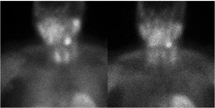
50 yo M with incidental elevated calcium, found to have elevated parathyroid hormone on further evaluation. Sestamibi scan was unrevealing. A 4D CT of the neck was performed.
Conventional early phase images were not very impressive. There were small lymph nodes noted.
On spectral analysis, a 1.1 cm nodule adjacent to the left lobe of the thyroid scan showed increased iodine uptake, and a distinct spectral curve, even though the attenuation was not very different from adjacent lymph node. A parathyroid adenoma was suspected.
On surgical evaluation, a thorough dissection of left level 4 was performed with multiple lymph nodes. Inferior to the lower pole of the left lobe of the thyroid gland, corresponding to the finding on CT, frozen section path came back as parathyroid tissue.
Intaoperative PTH fell from 83 to 35. Post-operatively, the PTH has remained stable at 42, and serum calcium is normal.
I believe it is time to retire the sestamibi scan for parathyroid imaging, and use 4D spectral CT.
Early and delayed sestamibi scan images are unremarkable. Uptake in left lower neck is in the submandibular gland.
Conventional CT image. Note left internal jugular vein (blue circle) and left common carotid artery (the big red). Yellow arrow points to parathyroid adenoma, and green arrow to an adjacent lymph node; both look nearly identical
On iodine map, the adenoma shows more uptake than the lymph node (2.1 mg/mL vs 1.2 mg/mL)
Attenuation curve of adenoma (magenta) and lymph node (blue) are quite different


