
Roses and islet cells
47 year old male with history of cirrhosis and gallstones presents with abdominal pain. Ultrasound showed a dilated common duct in addition to above findings. A surgical consult was obtained, given his medical conditions, and no convincing evidence of acute cholecystitis on ultrasound, GI consult was recommended.
GI team decided to do an endoscopy. And they found ulcers in the distal duodenum and proximal jejunum.
Now, everybody who has ever taken USMLE Step 1 knows that ulcers in this distribution mean that the patient has Zollinger-Ellison syndrome. Indeed his gastrin level came back markedly elevated at >600 pg/mL (normal is <100 pg/mL). But where is the gastrin-secreting primary? (for test-takers, the answer is that it is usually found in the so-called “gastrinoma traingle”)
Spectral CT to the rescue! He had a recent contrast enhanced scan, where the pancreas looked completely normal. But turn on spectral CT, and there is a clear nodule in the uncinate process that is consistent with an islet cell tumor!
Ga68 Dotate PET scan showed intense uptake in the nodule, consistent with neuroendocrine lesion.
This example again confirms utility of spectral CT in small pancreatic lesions, and with the detector based system, spectral CT is always on, and can be looked at in retrospect, and will reveal things that can be absolutely impossible to see on routine scans.
I looked up Dr. Zollinger and found “he was the president of almost every society he belonged to, including the American Board of Surgery, the American Surgical Association, the American College of Surgeons and even the American Rose Society.” Dr. Zollinger was also “feared by his students and loved by his patients. On rounds he was known to fire a resident on the elevator for some misdemeanor, only to rehire them by the time they had reached the 7th floor.”
Dr. Zollinger died of pancreas cancer in 1992.

Ulcer in second part of duodenum

Ulcer in third part of duodenum

Ulcer in jejunum

Conventional CT: Pancreas is NORMAL

Iodine map: Nodule in uncinate process (red arrow) with well-defined margin.

40 keV mono-energy image shows nodule well.
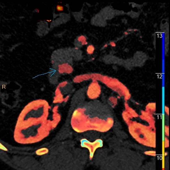
Z-effective map shows nodule shining (blue arrow)!
Ga68 Dotatate PET scan: Intense uptake in uncinate nodule. Note similarity to the Z-eff image above.

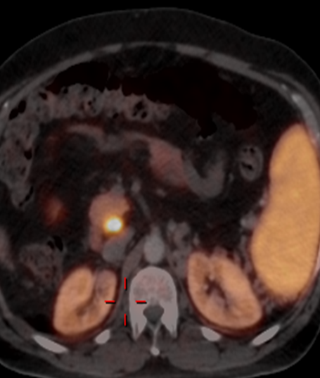
Ga68 Dotate PET scan whole body image. The intense uptake overlying the right kidney (red arrow) is the gastrinoma.