
Role of electron density weighted images
Case courtesy of Dr. Anthony Severt.
70 year old female presents to the hospital with fatigue, loss of appetite and abdominal pain for one month. She had subjective fevers, and white count was elevated. A non-contrast CT scan performed because of concern with renal function shows subtle hypodense areas in the liver, and portal vein thrombosis.
Turn on spectral CT, and electron density weighted images show the hypodense lesions much better! This is very interesting, because I thought that spectral CT was useful in contrast enhanced scans; but clearly, has a role in non-contrast scans as well!
Followup MRI shows the lesions very well, the T2W appearance and restricted diffusion is consistent with abscesses. Purulent material obtained by aspiration.

Non-contrast CT: Super subtle liver lesion in left lobe

Electron density weighted image: The left lobe lesion pops out. In addition, there are multiple abnormal foci in the right lobe that were simply not visible on the non-contrast image.
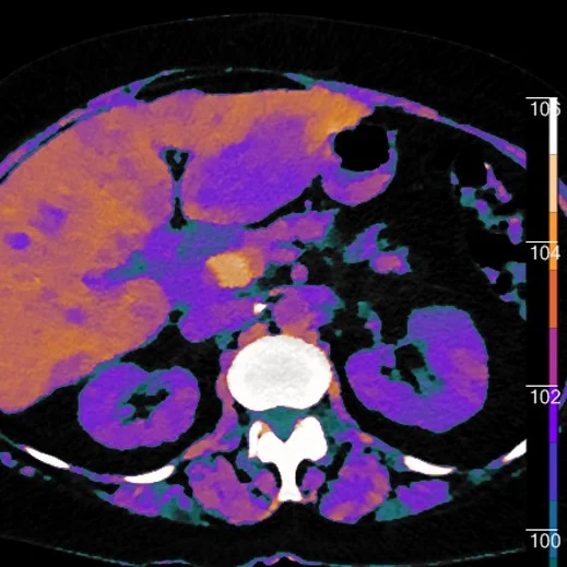
Color overlay with EDW image shows findings to good effect. Notice subtle areas of abnormality in the right lobe.

T2W MRI shows the left lobe of liver lesion as well as multiple areas of abnormality in the right lobe

DWI b=0 image

DWI b=800 image

ADC map. Note all abnormal areas have restricted diffusion. The left lobe lesion has a multiloculated appearance typical of abscess.