
Urinary stone composition
A 55 year old female presented to the ER with flank pain, nausea and vomiting. No fever, and labs are unremarkable. A routine CT performed with IV contrast shows a 2.5 mm obstructing stone in the left ureter. As the patient had severe pain, she was taken to the OR for a ureteroscopy with stone extraction.
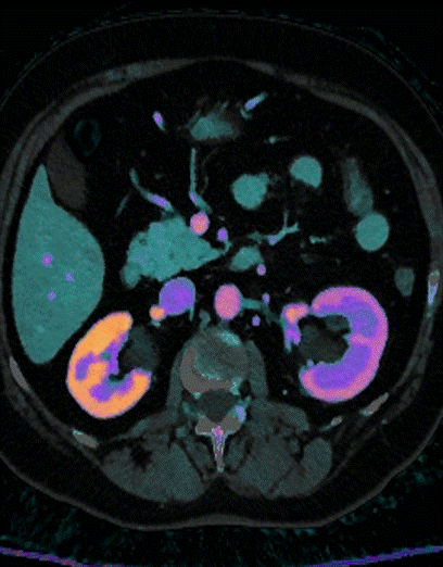
Can we tell more about the stone, than that there is a stone? Yes, of course, with Spectral CT! This calculus is made of uric acid on spectral analysis.
Stone was sent for analysis, which confirmed that it was made mostly of uric acid.
This is important, because it gives radiology a chance to go above and beyond the usual descriptions and into pathology, which is where we need to be. In this case, stone extraction was indicated because of unremitting symptoms, but in other case, a confident diagnosis of uric acid calculi may enable medical management and save the patient (and the healthcare system) procedures.
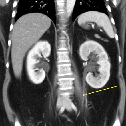
Routine conventional CT shows obstructing stone in left ureter (yellow arrow). There are peripelvic cysts in kidney bilaterally.
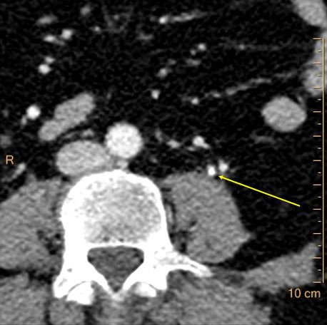
conventional CT (axial image). Stone in left ureter (yellow arrow)
Spectral CT, iodine overlay: Note delayed nephrogram in left kidney.
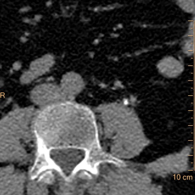
Virtual non-contrast: Stone pops out.
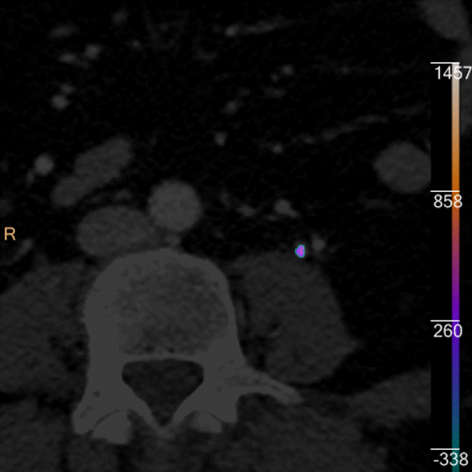
Uric acid overlay: Color coding of left ureteral stone confirms uric acid composition.

Morphological analysis after extraction: the stone is made of uric acid.