From Switzerland
Case and images are courtesy of Dr. Eva Geisler, radiologist at GZO Spital Wetzikon, near Zurich, in Switzerland. Dr. Geisler has been using Spectral CT for a while now, and like many of us, is blown away by the possibilities in daily diagnostic radiology. She sent me this case, and very graciously gave me permission to put up the images on this blog.
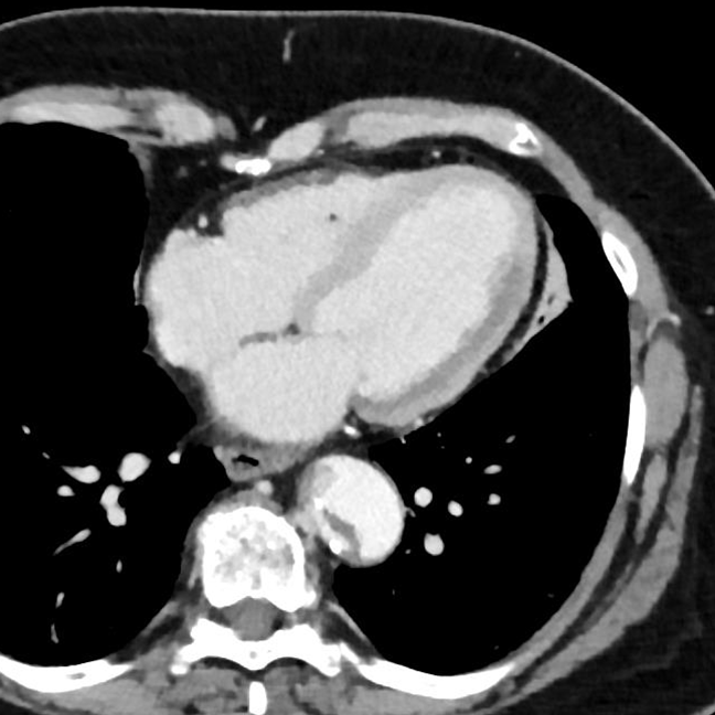
60 year old female presented to the hospital with chest pain. Venous phase CT shows a subtle area of decreased perfusion in the left ventricle. This is much more obvious on monoE images, and of course stands out on the iodine map and the Z-eff images. The arterial phase image shows the Stanford type A dissection, and involvement of the left main coronary artery.
These are spectacular images, and once again, highlight how with spectral CT, we can go above and beyond: talk not just about the primary pathology (the type A dissection in this case) but also, with a very high degree of confidence, we can talk about the complications.
Thank you, Dr. Geisler!
Conventional CT, venous phase. You might pick up hypoperfusion in lateral wall of left ventricle on a good day, but then again…
MonoE: Large perfusion defect is obvious!
Iodine overlay confirms perfusion defect
Z-eff image is a nice way to depict finding
Conventional CT, arterial phase shows Stanford type A dissection involving ostium of the left main coronary artery.


