The spleen is gone....believe me!
53 year old man brought in for intoxication. Blood alcohol level 0.301 g/dL. Blood pressure was soft, and free fluid was seen in the abdomen, so a CT scan was performed.
Obvious cirrhosis with large varices on CT. Spleen looks prominent, but otherwise not particularly noteworthy. Portal vein is patent. Small ascites.
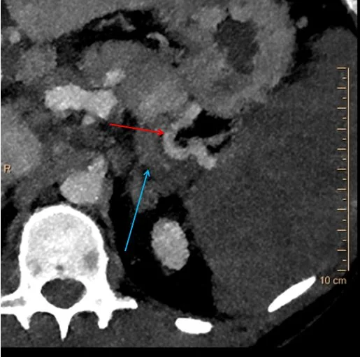
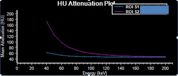
Turn on spectral, and something surprising shows up. The entire spleen has no iodine uptake. This is confirmed with a nearly flat spectral curve. On 40 keV images, the splenic artery is patent to the hilum, but the splenic vein appears thrombosed.
Focal splenic infarcts are pretty easy to call, with wedge shaped defects. A global splenic infarct is unusual, and can be difficult. Global spleen infarct from venous thrombosis is even more unusual. Iodine map makes the diagnosis very easy, and also make it easy to convince skeptics!
Conventional CT shows cirrhosis with varices. The spleen does not stand out as particularly abnormal.
Iodine map: The spleen is gone!
Iodine overlay: Oh so easy!
40 keV image: Splenic artery is patent to the hilum (red arrow). The splenic vein appears thrombosed (blue arrow). Note the spleen is very hypodense on the low keV image, consistent with absent iodine uptake.
Spectral curves: Blue curve (ROI in spleen) is nearly flat. Contrast with magenta curve (ROI in liver) which goes up on lower keV consistent with iodine uptake.


