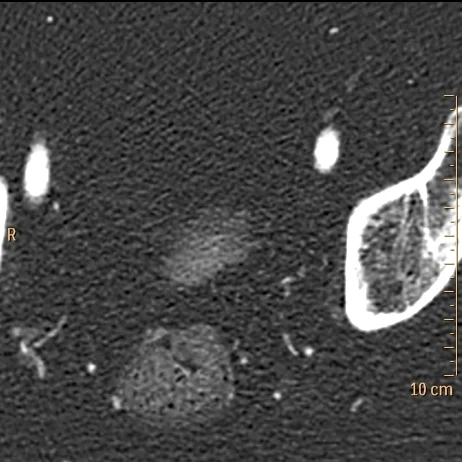
Bladder tumor
77 year old male on chronic antocoagulation presents with syncope. CT angiogram of the aorta was performed, and the aorta was without acute abnormalities. Pulmonary emboli were discovered.
Incidentally, a urinary bladder "filling defect" was discovered. On spectral analysis, there is intense uptake of iodine in the "filling defect", and it is therefore vascularized, and consistent with a bladder tumor.
Iodine maps can be very helpful when trying to differentiate hematomas from vascularized tissues.
Conventional CT shows a urinary bladder "filling defect"
Iodine map with intense uptake in the urinary bladder "filling defect", consistent with tumor
Perfusion nicely depicted on iodine overlay


