
Urothelial mass
71 yo M presented with acute onset left abdominal pain. He was previously scheduled for a urology appointment to work-up a urothelial mass, but came to the ED for acute onset pain.
Conventioanl CT shows a filling defect in the left renal collecting system. Iodine uptake seen on the fusion image confirms vascular lesion. Note the delayed nephrogram on the left consistent with obstruction from the mass.
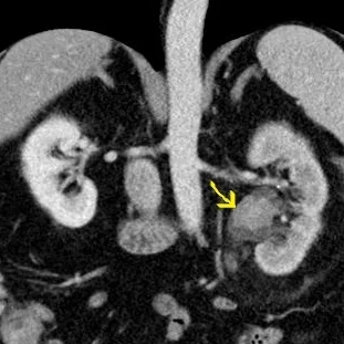
Conventional image with filling defect in left renal pelvis (yellow arrow)
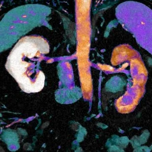
Iodine overlay with uptake confirms neoplasm
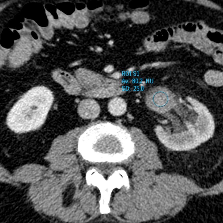

Region of interest placed on lesion shows spectral curve rising on lower keV, an elegant way to confirm iodine uptake and perfusion