
Pancreatic cancer
57 yo F presents with abdominal pain. Total bilirubin >6, Alkaline phosphatase >600. Routine CT obtained, shows biliary obstruction with nodule at the site of obstruction, possible stone or mass.
On spctral analysis, there is iodine uptake in the nodule obstructing the common bile duct, consistent with vascular tissue and excluding stone. Subsequent endoscopic ultrasound confirmed a pancreatic mass, and biopsy showed a pancreatic adenocarcinoma.

Coronal oblique conventioanl CT image shows biliary dilatation. Note hyperdense nodule (yellow arrow) obstructing the CBD.

Axial conventional image shows subtle nodule in pancreatic head

Nodule is much easier to see on 40 keV image

Fusion image shows clear iodine uptake in the nodule, consistent with tumor
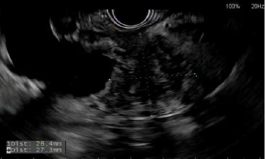
Image from endosopic ultrasound shows hypoechoic mass obstructing common bile duct