
Endoleak
Routine post-endograft placement CT scan, with increased density posterior to the graft
On virtual non-contrast, there is no density in same location
Color iodine overlay shows endoleak, probably type 2 endoleak from a lumbar artery
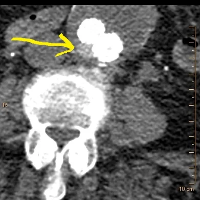
Conventional CT with focal hyperdensity adjacent to endograft
Virtual non-contrast
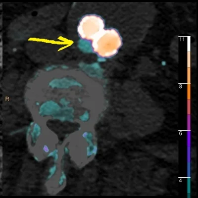
Fusion image nicely depicts the endoleak