
Myocardial ischemia at rest
84 year old male with history of pacemaker for sick sinus syndrome presents after syncope. Pacemaker interrogation shows ventricular tachycardia. A CT scan performed to evaluate for dissection shows a 4.9 cm abdominal aortic aneurysm.
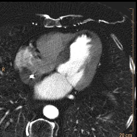
Closer look at spectral images shows hypo-perfusion in the left ventricular septum and apex. Subsequent coronary angiography showed severe 3-vessel disease with critical stenosis in the mid-LAD.
This patient would have received a cardiac workup regardless of spectral CT findings; nevertheless, this case illustrates how spectral CT can make obvious subtle findings that can be life-threatening.
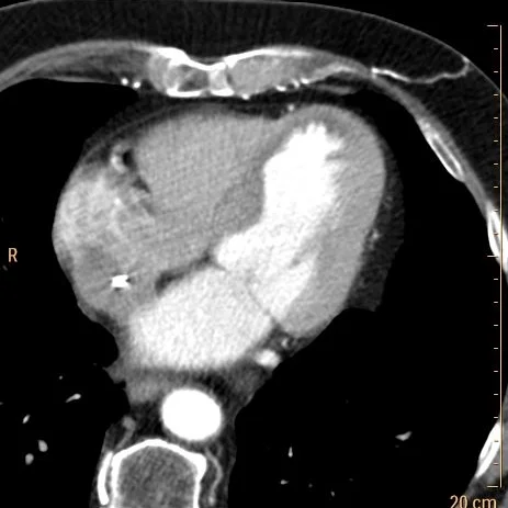
Conventional CT: The left ventricular myocardium looks not particularly interesting

Conventional CT: The septum shows slightly decreased attenuation, but hard to pick up from the noise
Iodine map: Clear decreased iodine uptake in septum (1.4 mg/mL vs 2.7 mg/mL)

Finding of septal hypoperfusion nicely depicted on iodine overlay

Spectral curves show significant difference between septum (blue) and lateral wall (yellow)
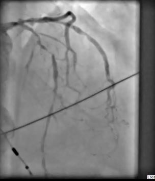
Coronary angiogram: Critical stenosis of the LAD. Severe 3-vessel disease