Optic nerve perfusion in CRAO
Full disclosure: I am a body imager, and this is the first neuroradiology case I am posting here, courtesy of my dear friend and colleague, Dr Ben Hoffman.
91 year old female presents with sudden painless loss of vision in the right eye. Fundoscopic exam consistent with central retinal artery occlusion. Head CT showed chronic left medial occipital infarct, and changes of small vessel disease. CT angiogram performed as part of stroke workup showed left PCA stenosis, ophthalmic arteries were patent.
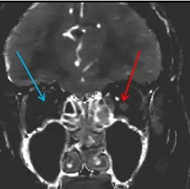
Dr Ben Hoffman turns on spectral. And lo and behold, the right optic nerve shows no iodine uptake. To the best of my knowledge, lack of perfusion on CT in CRAO has not been described before.
Patient was outside tPA window, and was treated with hyperbaric oxygen, unfortunately with minimal improvement.
Conventional CT, coronal plane. The optic nerves are unremarkable.
Iodine map: Absent uptake in right optic nerve (blue arrow). The left optic nerve shows good iodine uptake (red arrow).
Iodine overlay shows finding nicely!


