Goblet, anyone?
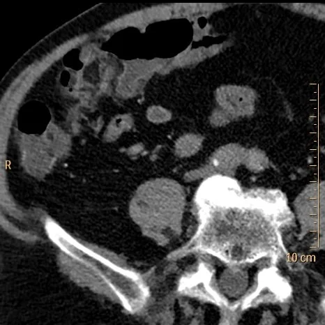
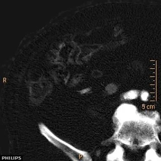
82 year old male with BPH presented to the ED with abdominal pain. Conventional single portal venous phase CT shows a “mass” in the right ureter. On spectral analysis, there is clear iodine uptake and enhancement of this mass, consistent with urothelial malignanacy.
Subsequent retrograde ureterogram shows a classic “goblet sign”. Biopsy confirmed a low grade papillary urothelial carcinoma.
Spectral CT certainly helped in this case, by confirming the enhancing nature of the lesion, but the main reason for putting this up is to show a great example of the goblet sign.
Per radiopedia, “goblet sign (or champagne glass sign) refers to the appearance of the ureter when it is focally dilated by an intraluminal mass. It is best seen when the ureter is opacified from below, by a retrograde ureterogram. Presence of this sign indicates the pathology to be chronic, permitting the lesion to be accommodated in the ureter.”
Conventional CT with filling defect in right ureter (yellow circle)
Virtual non-contrast: Comparison to conventional CT shows obvious enhancement visually. On measurement, HU increase from 36 to 82.
Iodine map shows clear iodine uptake, consistent with tumor.
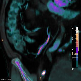
Iodine overlay nicely depicts the finding
Coronal oblique with iodine overlay: Note the ureteral dilatation above and below the enhancing mass.
And the Goblet sign!


