My oh MI!
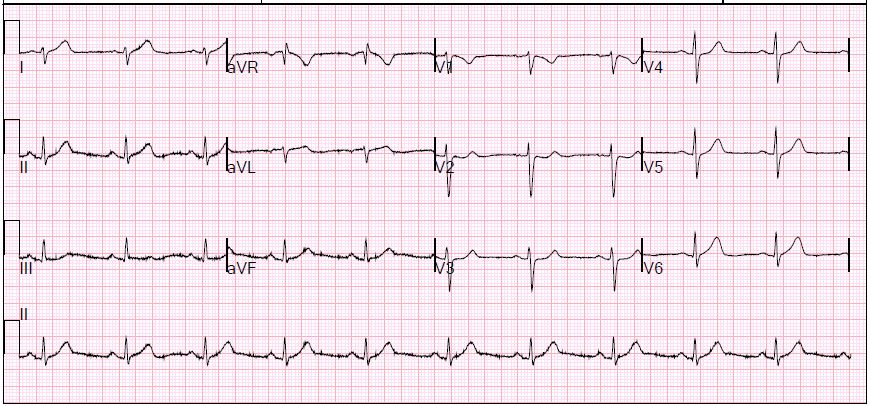
39 year old female with no prior history presents to the ED with right sided chest pain. Initial EKG with minimal ST-depression, and initial troponin elevated to 0.04, concerning for NSTEMI. Basic labs largely unremarkable (mild leukocytosis and hypokalemia). SL nitro did not provide any pain relief. Repeat EKG with dynamic changes-- resolution of ST depression. Delta troponin significantly increased to 1.04.
A CT angiogram of the aorta was performed to rule out dissection, and cardiology was consulted.
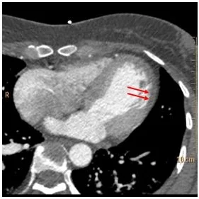
CTA shows no dissection. But look closely, and on the conventional images you can see relatively sharply defined endocardium in the lateral wall of the left ventricle, while the rest of the endocardium is fuzzy. I like this previously undescribed sign, it helps me detect cardiac hypoperfusion on non-gated scans.
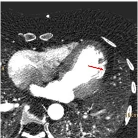
Now look at spectral images. There is clear iodine deficit in the lateral wall. This is well seen on the fusion image. This is an acute MI in the circumflex territory.
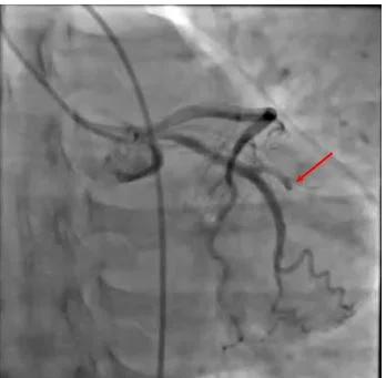
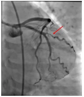
Occluded OM1, likely from a spontaneous dissection found in the cath lab and treated with angioplasty. Troponin peaked at 20. Patient discharged on hospital day 3, on plavix.
First EKG: Subtle ST segment changes, below the threshold of a radiologist
Second EKG: Dynamic ST changes. Wish I could tell you more.
Conventional CT: Focal sharply defined endocardium (red arrows). This is a very good sign for an acute MI on non-gated chest CT scans.
Iodine map: No iodine uptake consistent with hypoperfusion
Fused image with iodine overlay: Nice depiction of perfusion deficit
Cardiac cath: Focal occlusion of OM1, likely from a coronary dissection
Post angioplasty with good flow in OM1

