Another mass, another stone
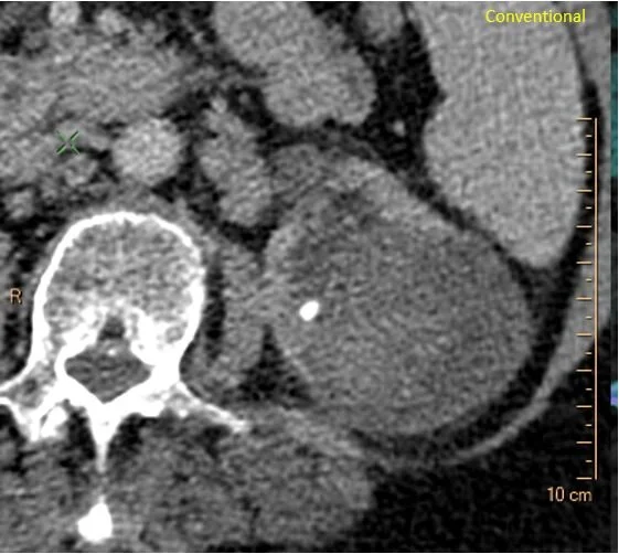
80 yo F presents to the ER with flank pain. CT scan of the abdomen is ordered. There is a complex heterogeneous mass in the left kidney, and a left kidney stone.
Now, at Hennepin, we do not like to recommend MRI till we absolutely have to. Instead, we turn on spectral CT, and we start looking for answers!
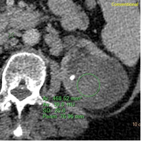
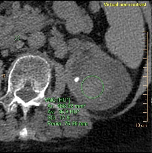
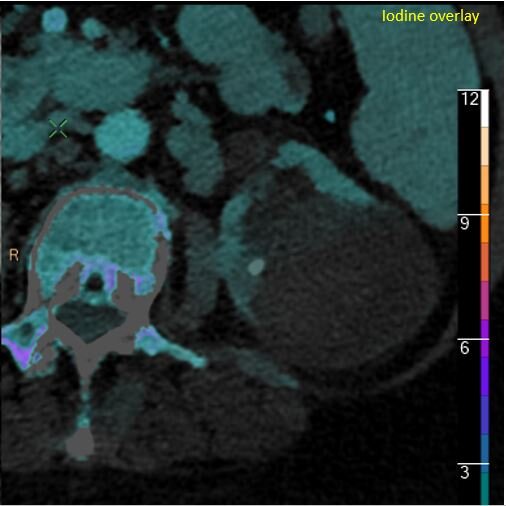
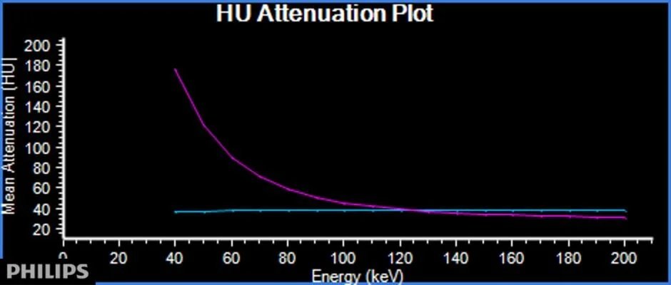
The mixed cystic and solid lesion has a solid component that is about 40 HU in density. On virtual non-contrast this is about 37 HU, so we can say that the solid component shows no enhancement. And there is no iodine uptake in the component. So this is hemorrhage in a cyst. Spectral curves prove this very elegantly.
But what about the stone you can see on the image?
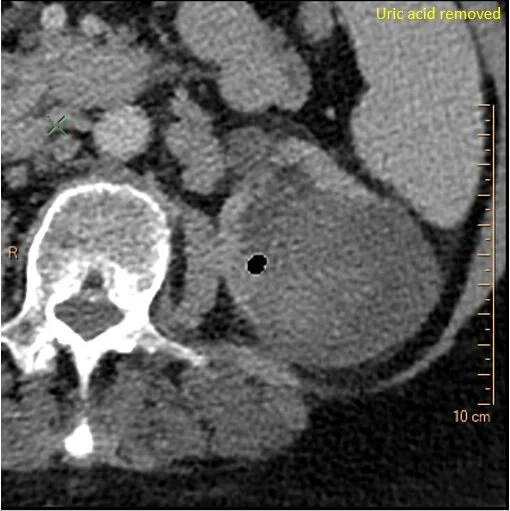
Spectral CT with uric acid removed shows the stone turns black, proving this is made of uric acid. This was confirmed after stone removal, on calculi analysis.
Conventional CT: Complex cystic and solid mass in left kidney and kidney stone.
Solid component is about 40 HU in density on conventional
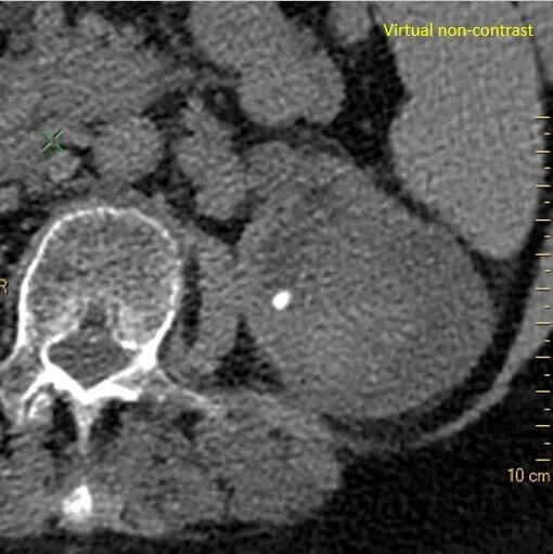
Virtual non-contrast: Solid component remains dense
Virtual non-contrast: Solid component is about 37 HU in density, proving no enhancement.
Iodine overlay: Absent iodine uptake proves this is not neoplastic.
Spectral curve: Blue curve on solid component remains flat. consistent with absent perfusion. Magenta curve, for comparison, is on perfused kidney parenchyma.
Uric acid removed: The stone is made of uric acid!

