
More about electron density
60 year old male with recently diagnosed metastatic melanoma presented for a routine CT abdomen and pelvis. On a chest CT done a day prior, he had extensive lung and mediastinal masses. The abdomen CT showed a small liver lesion, expertly picked up by the radiologist. The lesion is well seen on liver windows, and especially well seen on IMR (iterative model based reconstruction) images.
But can we make it stand out even more? With spectral CT, of course we can!
Usually my go-to to make lesion more conspicuous is the iodine map. In this case, the lesion is nearly imperceptible on the iodine map. Interestingly, the lesion is quite well seen on virtual non-contrast image.
Electron density weighted images compare the the electron density of the tissues to that of water (3.34 x 1029 electron x m3), expressed as a percentage. Electron density is the most important tissue property influencing photon and ion dose distributions in radiotherapy patients. Spectral CT can measure the electron density of tissues with a high degree of accuracy.
And electron density images can also make liver lesions stand out, as in this case, were the lesion in the right lobe just pops out. Corresponding MRI and PET images confirm that it is a malignant lesion.
To the best of my knowledge, there are no studies looking at liver lesion detection with electron density weighted images. This blog is where you read about this first!
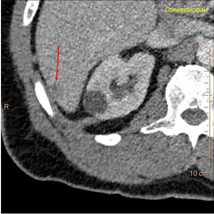
Conventional CT: The lesion in the liver is very hard to see.
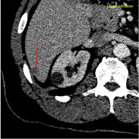
Liver windows make the lesion more conspicuous.

IMR, with it’s noise reducing ability, makes the lesion impossible to miss.
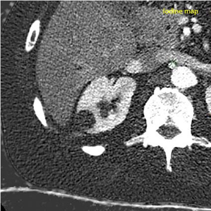
Iodine map: The lesion is not visible! Has same iodine uptake as normal liver. This is a portal venous phase image.
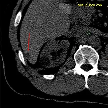
Virtual non-contrast: Lesion pops out again.
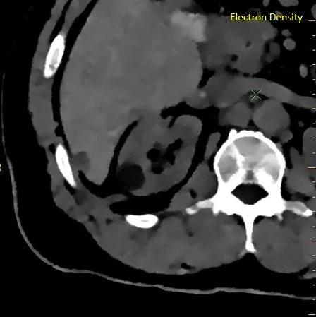
The best contrast resolution, by far, is on electron density image!
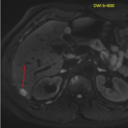
MRI with DWI showing lesion.
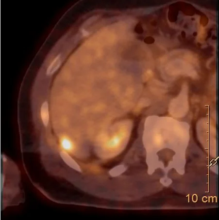
Hypermetabolic on PET/CT.