
A case of rectal bleeding
70 year old female, postop day 20 status post sigmoid resection and anastomosis, presents with acute onset of rectal bleeding. A routine single phase CT performed in the ER, which is interesting, since we have a specific GI bleed protocol which has a non-contrast scan, followed arterial and portal venous phase scans.
With spectral CT, do we need a multiphasic phase, really? Maybe not…
In this case, see how the virtual non-contrast and iodine maps easily delineate all the important features: there is active bleeding arising from near the sigmoid anastomosis. This was confirmed with flex sig, where the bleeding vessel was found and successfully treated with clipping.

Conventional CT: Red arrow shows “hyperdensity” in the rectal lumen.
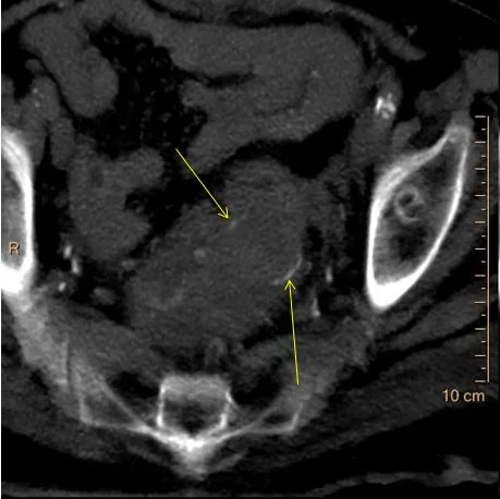
Virtual non-contrast: The “hyperdensity” is gone! So you do not really need a true non-contrast exam. The yellow arrows show the anastomosis.

Iodine map: Confirms bleeder arising from near the anastomosis
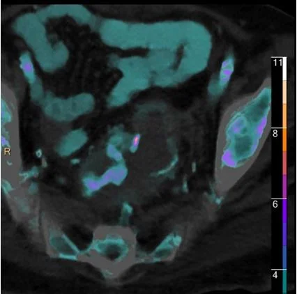
Iodine overlay: Nicely confirms findings

Bleeding vessel seen on flexible sigmoidoscopy
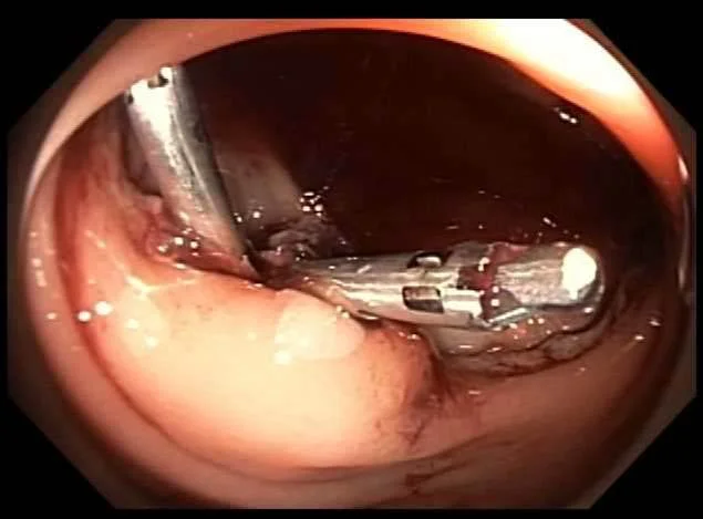
Clips placed successfully.