
Lightbulb sign...
61 yo M presents with weight loss and anemia, hemoglobin of 4 mg/dL. Received 2 units of blood in the ED, and routine abdominal CT shows colon mass, and incidental aortic aneurysm. Pancreas reportedly normal.
On subsequent review, a subtle nodule is visible in the neck of the pancreas adjacent to the SMV. This is much better seen on 40 keV image and iodine map. Virtual non-contrast confirms lack of calcification in nodule.
Intraoperative ultrasound confirmed nodule, and after discussion with patient, a conservative course of action was adopted.
40 keV images can make pancreatic islet cell tumors (and any iodine avid lesion) shine like a lightbulb!

Conventional CT shows mass in cecum. Note incidental AAA.

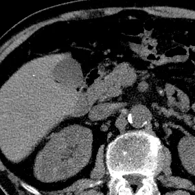
Axial image shows very subtle lesion in neck of the pancreas

40 keV mono-energy image shows nodule in pancreas much more clearly. Lightbulb!

Iodine map with uptake in nodule
No calcification seen on virtual non-contrast image.