LV aneurysm with thrombus
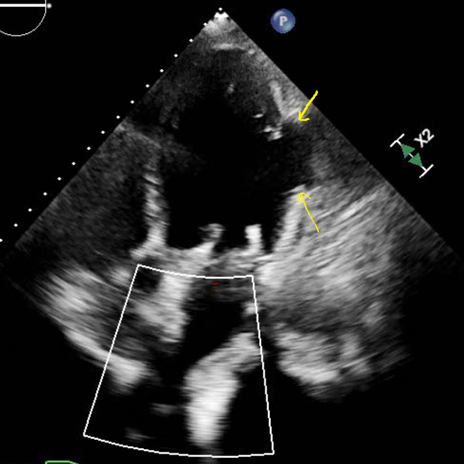
69 yo F with previous NSTEMI and ischemic cardiomyopathy, presenting with chest pain and shortness of breath. Conventional CT shows outpouching of the lateral wall of the left ventricle.
On spectral analysis, there is absent iodine uptake in wall of aneurysm with large filling defect consistent with thrombus. This is well seen on fusion image.
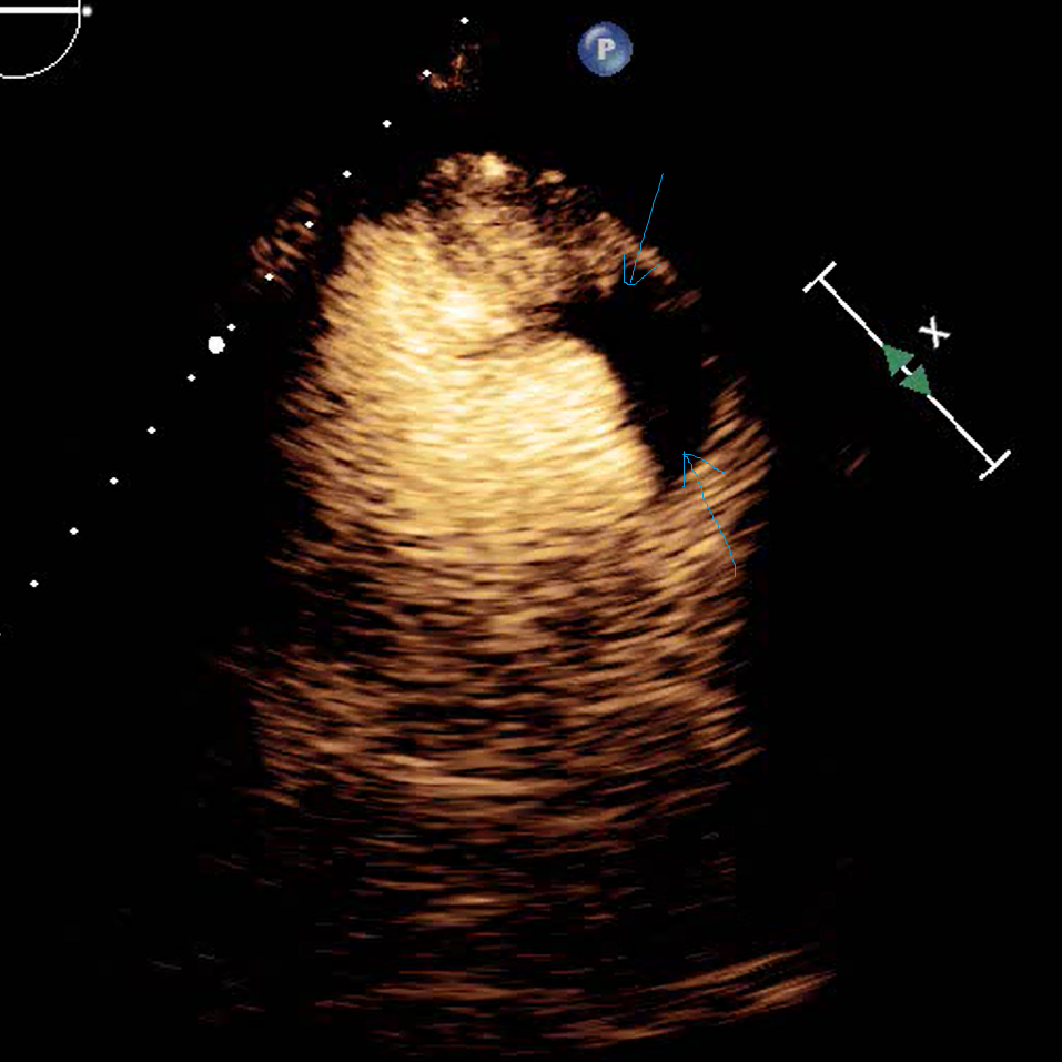
Echocardiogram with contrast confirms findings. Patient started on anticoagulation.
Conventional CT shows large left ventricle lateral wall aneurysm (yellow arrows)
Iodine map shows large thrombus in the aneurysm with absent iodine uptake (red curve)
Iodine overlay image. Note normal LV wall perfusion (white arrow), color is absent in the aneurysm.
Echocardiogram (HLA view) shows obvious lateral wall aneurysm (yellow arrows)
Large echolucent area (blue arrows) after contrast confirms thrombus


