
Internal hernia with necrotic colon
55 yo F with abdominal distention and vomiting. Conventional CT shows distended and malpositioned cecum and ascending colon, with a mesenteric swirl. On spectral analysis, note lack of perfusion in the distended colon and terminal ileum.
Lactate came back at 11. Patient taken emergently to the OR. Internal hernia with cecum and right colon herniated behind a Billroth 2 gastrojejunostomy. Colon necrotic and foul smelling, partial colectomy performed.
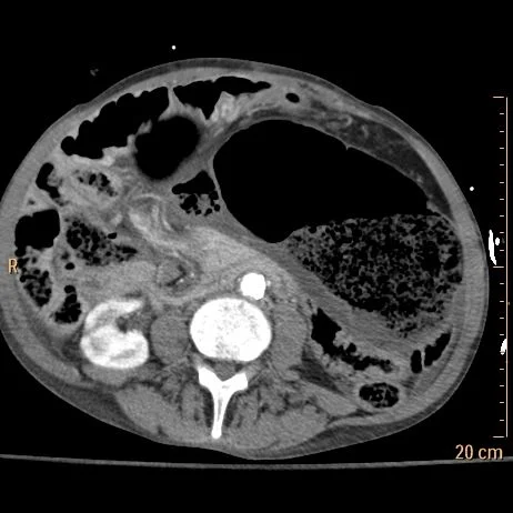
Conventional CT with dilated malpositioned cecum

Iodine map with no perfusion in wall of cecum and terminal ileum
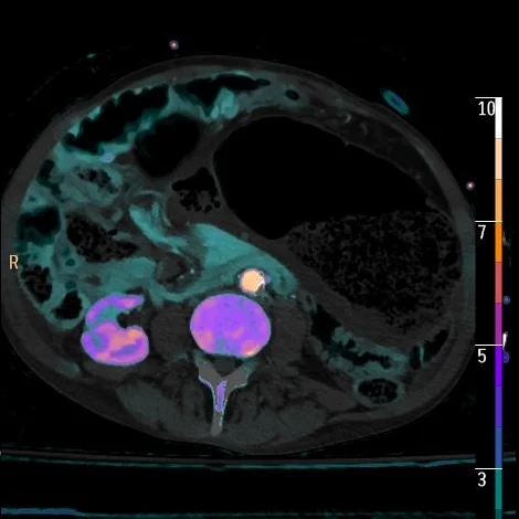
Iodine density overlay with no perfusion in wall of cecum and terminal ileum

Dilated segment of colon extends into pelvis, note absent perfusion in the wall on overlay image

Intraop image courtesy Dr Ryan Fey; dilated necrotic colon confirmed