
Painless hematuria
77 year old male presents to the ED with painless hematuria. A routine CT scan performed, shows a filling defect in the urinary bladder, with a linear calcification. Now is that a mass?
Spectral to the rescue! The filling defect shows no iodine uptake, and is increased density on virtual non-contrast image. The spectral curve is flat, consistent with lack of iodine uptake. This is a bladder clot.
My super-smart resident does a prone image and the filling defect is clearly mobile, confirming what spectral told us.

Conventional CT: Filling defect posterior urinary bladder with linear calcification. Also note the right sided bladder diverticulum with a dependent stone.

Virtual non-contrast: The filling defect is hyperdense.

Iodine map: No iodine uptake in filling defect. This is a clot around a bladder stone.
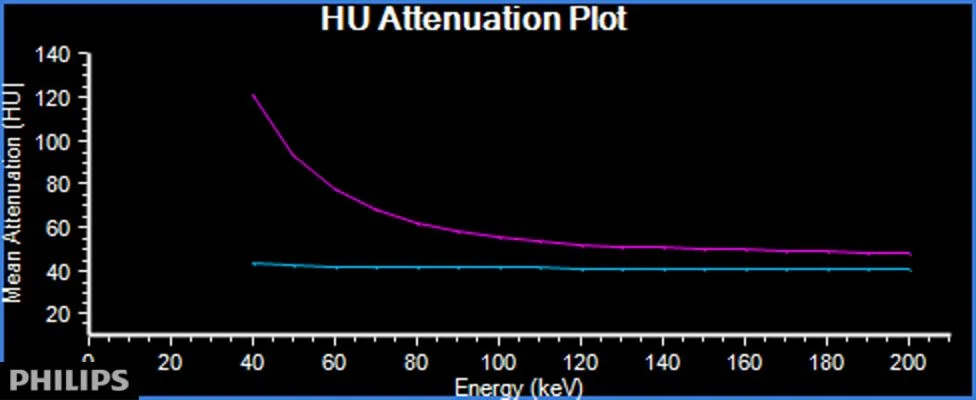
Spectral curve: Blue line in clot in flat. Magenta line (in left femoral vein) for comparison. Iodine containing ROI will show increase in attenuation on lower energies.

Prone conventional CT (delayed): The clot has moved to the dependent bladder!