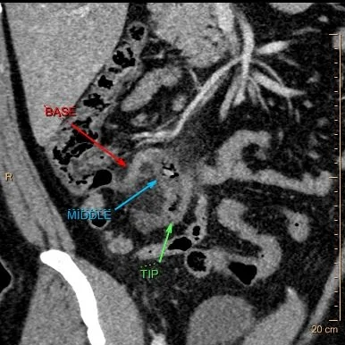
Does this appendix come out?
38 year old male presents to the ER for abdominal pain for the last 24 hours. WBC count was 9.77 k/cmm. CT scan was performed, shows clear acute appendicitis. Patient was taken to the OR, where appendicitis was confirmed. There was purulent fluid around the appendix.
So can we diagnose perforation on CT?
This paper from Philadelphia, published in 2003, talks about discontinuous enhancement as a very good sign of appendiceal perforation on CT. Over the years, I have found it very accurate. But it is a sign that takes experience to recognize, and may be difficult to get residents to appreciate. And spectral CT, of course, can help!
Going back to the CT scan on our patient, note the base of the appendix is normal, and there is a lot of inflammation around the middle of the appendix, where there is a small appendicolith. There is markedly decreased iodine uptake in the wall, and on the iodine map, there is no uptake where the normal wall should be. This is a clearly perforated appendix!
Path report : "Pink-tan to hemorrhagic and rough with an area of perforation in the middle appendix."
Now, why is it important to diagnose this pre-operatively? Well, I am no surgeon, but there is some controversy about whether a patient with a perforated appendix (w/o an abscess, as in this case) should be taken to the OR, or treated conservatively with a course of antibiotics first. The chances of a post-op abscess if the patient is taken to an immediate appendectomy is pretty high, at least 20-30% in my experience. Regardless how these patients are managed at your institution, it is very important to give the surgeon the best possible information.
Our patient returned 10 days later with a RLQ abscess that needed a surgical drain.
Conventional CT: Note obviously inflammed mid-appendix with small lith.
Iodine map: The wall of the middle of the appendix is missing! You can again make out the wall in the distal appendix.
40 keV mono-energy image shows finding very well
Conventional CT with iodine overlay: Now I am just showing off, but the perfortaion is obvious!
10 days post-op: Large RLQ abscess. This was managed with a drain, that stayed in for a month.


