A marginal ulcer
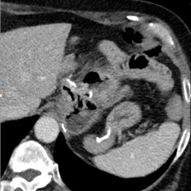
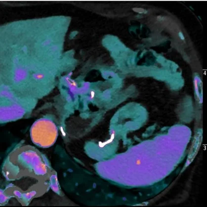
53 year old female with prior Roux-en-Y gastric bypass (6 years ago) and ongoing tobacco use presents with abdominal pain and melena. Hemoglobin came back at 9.5 mg/dL. A CT scan was obtained. Conventional images show subtle abnormality at the gastrojejunostomy site. Iodine map shows clear large defect in wall enhancement adjacent to the GJ anastomosis, consistent with an ulcer.
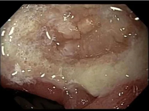
Subsequent endoscopy confirmed large marginal ulcer. Patient discharged on conservative management.
Marginal ulcers are a relatively common complication of bariatric surgery, and may lead to catastrophic bleeding. A large residual parietal cell mass is thought to contribute. As in this case, the added advantage of spectral CT in recognizing wall abnormalities in the GI tract can make diagnosis very easy.
Conventional CT. Staple line indicates the gastric bypass.
Iodine map: The wall discontinuity from the marginal ulcer (orange arrow) is obvious.
Iodine overlay shows finding well
Image from endoscopy shows large ulcer at the GJ anastomosis
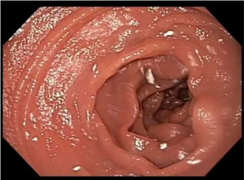
Normal mucosa distal to the GJ anastomosis


