
Hyperdense ascites
Case courtesy of Dr. Hoffman and Dr. Pogatchnik.
60 year old inpatient undergoes a CT of the abdomen and pelvis for persistent lactacidosis. IV contrast not given as he is in acute renal failure (eGFR 20 mg/mL/1.73m2). There is a moderate amount of ascites, which is hyperdense, with attenuation of 20-35 HU. So is it blood?
The rad resident, and my colleague reading the scan, are well versed in the use of spectral, and look to spectral for an answer.
28 hours prior, patient had a CT of the chest, abdomen and pelvis for trauma, and had received contrast at the time. On the current scan, note the persistent nephrogram (consistent with ATN), vicarious excretion in the gallbladder, and hyperdensity in the colon. Iodine map shows clear uptake of iodine in ascites. On virtual non-contrast, the ascitic fluid is low density (0-5 HU).
These findings are consistent with vicarious excretion of iodinated contrast in ascites in a patient with renal failure. This is a well-known phenomenon, described in this beautiful paper in 2009 in the AJR from UCSF. While they inferred the phenomenon, we can prove it using spectral CT!
Personally I find this to be the most exciting application of spectral CT: furthering our understanding of physiologic processes (as in this previous case).
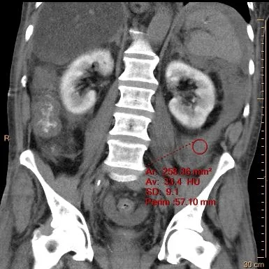
Conventional CT: coronal plane. Persistent nephrogram 28 hours after contrast admin. There is hyperdense ascites in left pericolonic gutter (33 HU).

Virtual non-contrast: The ascitic fluid is low density (0.9 HU).

Iodine map: Clear iodine uptake in ascitic fluid.

Fused axial image with iodine overlay shows iodine uptake in perisplenic fluid, and excretion in the gallbladder.

Fused axial image with iodine overlay shows iodine uptake in pericolonic fluid, as well as iodine excretion in the colon (this may be from biliary excretion or direct GI excretion of contrast).