Of course, we can do gout
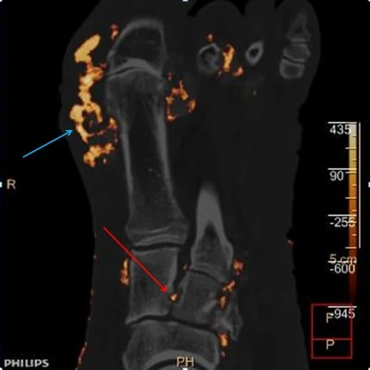
55 year old male presents to the ED for a foot ulcer. He has an aversion to medical care, has not seen a doctor for years, and does not take any medications. On physical exam, a large ulcer overlies the first MTP joint. CT scan was performed, showing extensive erosive arthritis and multiple soft tissue tophi.
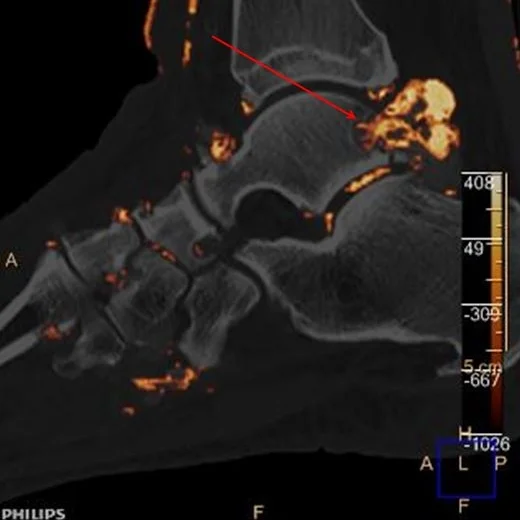
Material decomposition with spectral CT can identify composition of tophi. In this case, urate deposition was confirmed. Identifying tissue composition is one of the most elegant uses of spectral CT. Used in this way, many more deposits of uric acid can be identified.
A long-standing history of untreated gout was elicited.
Volume rendered CT image: Large ulcer over the first MTP joint
CT with uric acid overlay: Large urate tophus over the first MTP joint (blue arrow). Note suble erosion between 1st and 2nd cuneiform with uric acid (red arrow)
CT with uric acid overlay: Anterior and posterior tendons with urate deposition (blue arrows). Note urate deposition in subtalar joint (red arrow)
CT with uric acid overlay: Large tophus eroding into the talus (red arrow)


