
A case of focal infiltrate
Young female with pleuritic chest pain presented to the ED. A PE study was obtained. A right lung wedge shaped pleural based opacity was seen, with mediastinal adenopathy. On spectral analysis, the pleural based opacity has decreased perfusion compared to normal lung, and the lymph nodes also appear hypoperfused (caveat: this may relate to the phase of the scan).
As malignancy was suspected, a PET scan was obtained, which shows marked FDG uptake in the lung opacity and lymph nodes. EUS guided FNA shows necrotizing granulomas, and serologic tests were consistent with histoplasmosis.
Many patients with chest symptoms get CT scans, and in my experience, decreased perfusion in lung opacities is often seen in infectious conditions. While more study is needed, spectral imaging can be a useful adjunct in diagnosing these often difficult cases.
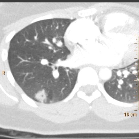
Conventional CT with pleural based opacity in right lung. Astute obervers will notice small cavitation.

Notice focal hypoperfusion in the lung opacity and adjacent air-trapping.
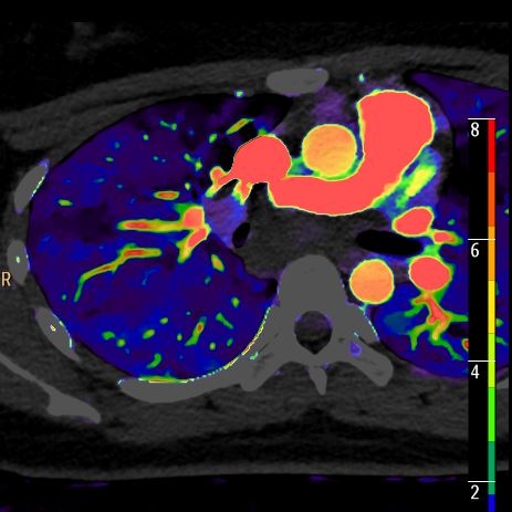
Large subcarinal lymph node with decreased perfusion, suggesting necrosis
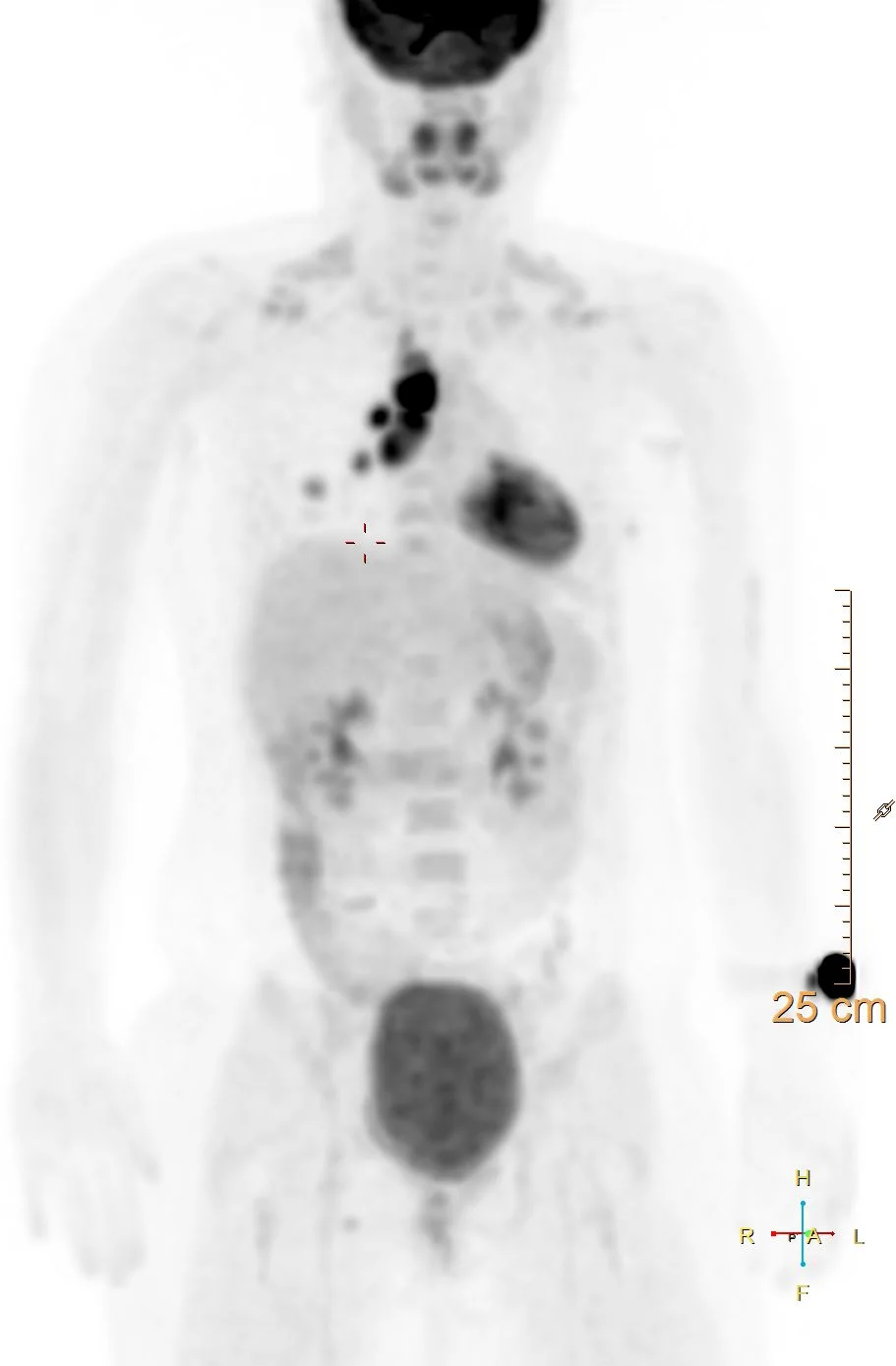
intense FDG uptake in mediastinal lymph nodes and lung opacity.